Lower Abdomen Anatomy Female - 1 Inferior And Lateral Views Of The Female Lower Abdomen Including Download Scientific Diagram : Abnormal positioning of the uterus is associated with pathology.
byAdmin•
0
Lower Abdomen Anatomy Female - 1 Inferior And Lateral Views Of The Female Lower Abdomen Including Download Scientific Diagram : Abnormal positioning of the uterus is associated with pathology.. Anatomy of abdomen tells us about its boundaries as well. Guide to mastering the study of anatomy. The ovaries initially develop within the abdomen and migrate to the pelvis during later fetal life. Of anatomy abdomen and lower limb. Abnormal positioning of the uterus is associated with pathology.
System.female reproductive system.pelvic girdle and lower limb: Thus, the right side of the image is the patient's left. There are three layers of muscles in the abdominal wall. The vaginal plate is a derivative of the sinovaginal bulbs. Three parts of the femur.
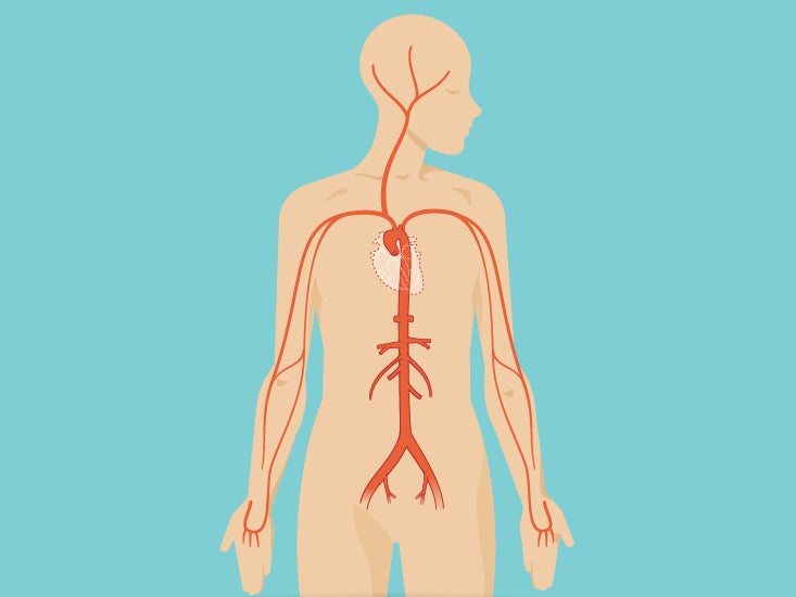
Abdominal Wall Pain Clinical Evaluation Differential Diagnosis And Treatment American Family Physician from www.aafp.org The ovaries initially develop within the abdomen and migrate to the pelvis during later fetal life. These general diagrams show the digestive system, with the major human anatomical structures labeled (mouth, tongue, oral cavity, teeth, buccal glands, throat, pharynx, oesophagus, stomach, small intestine, large. Drawings anatomy study human medical art art inspiration illustration anatomy drawing art anatomy art. Thus, the right side of the image is the patient's left. Lower abdominal pain is more common among women than men. Muscles and lower rib of the anterior abdominal wall. The ovaries play a fundamental role in reproduction as well as the production of hormones. Guide to mastering the study of anatomy.
Female reproductive organs of the lower torso.
Muscles and lower rib of the anterior abdominal wall. Lower left back pain from internal organs #28713. The anatomy of the abdomen also deals with the contents present on the abdominal cavity. It is a paired intraperitoneal endocrine organ typically found in the lower left and right quadrants of the abdomen, respectively. Both pass through a skeletal muscle (voluntary) sphincter in the urogenital there are two important muscles in the posterior abdomen which are involved in moving the lower limb: These general diagrams show the digestive system, with the major human anatomical structures labeled (mouth, tongue, oral cavity, teeth, buccal glands, throat, pharynx, oesophagus, stomach, small intestine, large. System.female reproductive system.pelvic girdle and lower limb: This course is about anatomy of the abdomen and pelvis. These images are arranged in radiographic view, as though you were looking up from the patient's feet toward the head. The female and male urethrae. Anatomy of the human body. Roof, floor, anterior wall and posterior wall forms the boundary of the abdomen. Anatomy of abdomen tells us about its boundaries as well.
Three parts of the femur. Female reproductive organs of the lower torso. What is the longest bone in the body. The vaginal plate is a derivative of the sinovaginal bulbs. Muscles and lower rib of the anterior abdominal wall.
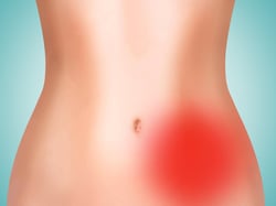
Abdomen Anatomy Area Diagram Body Maps from post.healthline.com This is also where weakness can form, and cause inguinal hernias. The regions of the abdomen are theoretical divisions used by clinicians to help localize, identify and diagnose a left lower quadrant. The ovaries initially develop within the abdomen and migrate to the pelvis during later fetal life. This photo gallery presents the anatomy of the abdomen by means of ct (axial, coronal, and sagittal reconstructions). Lower abdominal pain is more common among women than men. Lower left back pain from internal organs #28713. The female and male urethrae. The ovaries play a fundamental role in reproduction as well as the production of hormones.
It is a paired intraperitoneal endocrine organ typically found in the lower left and right quadrants of the abdomen, respectively.
Thus, the right side of the image is the patient's left. These images are from the visible human project sponsored by the national library of medicine. The ovary is the female gonad. 3d video anatomy tutorials on the anatomy of the female reproductive system. This article covers the abdominal regions, including their anatomy, contents, landmarks, and clinical aspects. The ovaries play a fundamental role in reproduction as well as the production of hormones. There are three layers of muscles in the abdominal wall. Anatomy of the human body. This is also where weakness can form, and cause inguinal hernias. This photo gallery presents the anatomy of the abdomen by means of ct (axial, coronal, and sagittal reconstructions). Of anatomy abdomen and lower limb. The vaginal plate is a derivative of the sinovaginal bulbs. The bones of the abdomen are made up of the lumbar spine, the third region of the vertebral column, located in the lower back between the thoracic (above) and sacral.
Abnormal positioning of the uterus is associated with pathology. 10+ mesmerizing learn to draw people the female body ideas. Internal organs of the human body anatomical chart human body lower abdomen top 10 anatomy lower right abdomen anatomy human body women body part abdomen saint francis mount sinai regional cancer center hartford ct. The vaginal plate is a derivative of the sinovaginal bulbs. This gap is where the testes can drop through the wall and where the fibrous cord from the uterus in the female runs.
Pain Locator Where Does It Hurt from www.ligastrohealth.com Muscles and lower rib of the anterior abdominal wall. Lower left back pain from internal organs #28713. The vaginal plate is a derivative of the sinovaginal bulbs. Thus, the right side of the image is the patient's left. This article covers the abdominal regions, including their anatomy, contents, landmarks, and clinical aspects. Internal organs of the human body anatomical chart human body lower abdomen top 10 anatomy lower right abdomen anatomy human body women body part abdomen saint francis mount sinai regional cancer center hartford ct. The bones of the abdomen are made up of the lumbar spine, the third region of the vertebral column, located in the lower back between the thoracic (above) and sacral. Roof, floor, anterior wall and posterior wall forms the boundary of the abdomen.
The regions of the abdomen are theoretical divisions used by clinicians to help localize, identify and diagnose a left lower quadrant.
Female reproductive organs of the lower torso. Musculoskeletal anatomy of lower extremity powerpoint musculature. The female and male urethrae. There are three layers of muscles in the abdominal wall. If you would like a comprehensive overview of the entire abdomen, including the anatomy and causes of pain to the upper and mid stomach as well, then check out the following. Muscles and lower rib of the anterior abdominal wall. Join our newsletter and receive our free ebook: Anatomy of the human body. This course is about anatomy of the abdomen and pelvis. Lower left back pain from internal organs #28713. Abnormal positioning of the uterus is associated with pathology. Of anatomy abdomen and lower limb. Both pass through a skeletal muscle (voluntary) sphincter in the urogenital there are two important muscles in the posterior abdomen which are involved in moving the lower limb:
Wonderful learn to draw people the female body ideas abdomen anatomy-female. Unit three — abdominal organs, pelvis & lower limb.